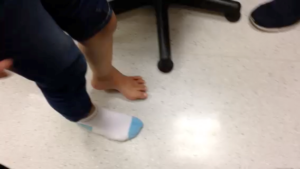
What is Cavus Foot?
The Cavus Foot refers to an abnormal elevation of the medial longitudinal arch of the weight-bearing foot. A High Arch Foot does not necessarily always mean it is a Cavus Foot, as a Cavus foot denotes an underlying Cavovarus or Calcaneovalgus type foot.
A High Arch Foot can merely be a normal variant. However, like the flat foot, it can also sometimes be symptomatic.
What are the causes and symptoms of cavus foot?
The Cavovarus Foot, however, needs a closer looking-at. Approximately 2/3 are associated with an underlying neurological condition. When it occurs in both feet it tends to denote a hereditary or congenital (present from birth) component.
The Most Common cause in a bilateral Cavovarus Foot is Charcot-Marie-Tooth (CMT) disease. CMT is a hereditary sensorimotor neuropathy that leads to muscle weakness and sensory changes. It is the muscle imbalance that leads to the Cavovarus deformity.
In the unilateral Cavovarus foot, one must rule out lesions in the spinal cord. Other causes may include a previous fracture which healed in malunion or may be Idiopathic, in which no known cause is found.
How is cavus foot diagnosed?
In the Cavovarus Foot, the 1st Ray (Big toe) is plantarflexed (lowered), and the forefoot (front part of foot) is adducted and in pronation (turned inwards and downwards). This is due to a weakness in the peroneus brevis, and a stronger peroneus longus; as well as a strong pull of the tibilais posterior muscle.
The Hindfoot (back part of foot) goes inwards (varus) as a consequence in order to balance and form a tripod with the forefoot. As time progresses the hindfoot becomes stiff in this position.
This deformity leads to instability during walking, ankle sprains, painful callosities under the heads of the toes, clawing of toes. As time progresses, as there are abnormal articulation and pressure on the joints, arthritis may set in.
Assessment begins with the ruling out of any possible neurological condition or spinal cord abnormality. This may require a referral to a neurologist and the need for other imaging. Once these have been addressed, we can then focus on the deformity afflicting the foot.
X-rays are performed and the foot is assessed to see which muscles and nerves are functioning well or affected. Joints are assessed to see if they are flexible.
A Coleman block test is a simple clinical test that can be performed in the clinic to assess if the hindfoot is fixed or flexible and aids in planning if surgical intervention is warranted. CT scans and Nerve conduction studies may be performed if surgery is being planned for.
How is cavus foot treated?
Conservative treatment is always attempted first, as with most conditions. This entails footwear adaptation, ankle support, physiotherapy to strengthen the weak muscles and stretch the overpowering ones.
Orthoses may be used to prevent contractures, callosities as well as to make the gait more energy efficient.
If conservative treatment fails, or if the foot is rigid in its deformity, surgery is indicated in order to alleviate the pain and symptoms and to allow the patient to function as normal as possible, with a plantigrade (neutral) foot.
The surgery that is performed is contingent on the flexibility of the foot and its joints. Nerve conduction studies performed will tell us which muscles are functioning. Their tendons may be transferred in a way to help correct the deformity and function.
Bone Cuts (osteotomies) may also be performed in order to aid in the alteration of biomechanics. The fusion of joints may be required where joints are rigid or arthritic.
This is avoided as much as possible in the paediatric patient and indicated more in the adult cavovarus foot.
The circular ring fixator is a very powerful tool which can be used in the correction of these deformities. It stands to reason that since these deformities are longstanding, they may not be corrected overnight with a single-stage surgery.
The circular ring fixator (Ilizarov) technique allows the correction to be gradual and over a longer course of time.

Looking For A Reliable Paediatric Orthopaedic Orthopaedic Specialist?
Fast Medical Attention, Transparent Fees
Make an appointment for comprehensive care for your paediatric orthopaedic problems!
