Introduction to your Spine
Understanding your spine and how it works can help you better understand the conditions that occur from wear and tear such as neck pain and back pain, as well as other spine problems such as scoliosis and sciatica.
The spine functions to hold up your head, shoulders, and body. It gives you support to stand up straight, and it also gives you flexibility to bend and twist the neck and back. It also protects your spinal cord. The spinal cord transmits nerves from the brain to their destinations e.g. extremities and various organs of the body.
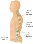
Your spine is made up of three segments, i.e neck (cervical spine), upper and middle back (thoracic spine), and the lower back (lumbar spine). When seen from the side, these segments form three natural curves (Figure 1). The “c-shaped” curves of the neck (cervical spine) and lower back (lumbar spine) are called lordosis. The “reverse c-shaped” curve of the upper and middle back (thoracic spine) is called kyphosis.
These curves are important to balance and they help us to stand upright. There is a range of normal values for each of the natural curves, and we often see loss of the normal c shaped curves of the neck (cervical spine) and lower back (lumbar spine) as part of wear and tear of the spine. If any one of the curves becomes too large or small, it becomes difficult to stand up straight and our posture appears abnormal.
Abnormal curvatures of the spine are also referred to as spinal deformity. These types of conditions include kyphosis of the thoracic spine (“hunchback”) and loss of lordosis of the lumbar spine (“flatback”).
These normal curves when viewed from the side should not be confused with spine scoliosis, which is an abnormal curve when seen from the back, and not the side. Scoliosis is a sideways curvature that makes the spine look more like an “S” or a “C”.
Structure
The spine is made up of small bones, called vertebrae, which are stacked on top of one another and create the natural curves of your back. The vertebrae are not in direct contact with one another, but kept apart by the intervertebral discs, which are sandwiched between the vertebrae.
Vertebrae
These bones connect to create a canal that protects the spinal cord (Figure 2). The cervical spine is made up of seven small vertebrae that begin at the base of the skull and end at the upper chest. The thoracic spine is made up of 12 vertebrae that start from the upper chest, and are connected to the rib cage. The lumbar spine consists of five larger vertebrae. These vertebrae are larger because they carry more of your body’s weight. The sacrum and coccyx (tail bone) form the bottom most portion of the spine, and they are joined to the pelvis via the sacro-iliac joints.
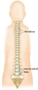
Figure 2 Image courtesy of AAOS (American Academy of Orthopaedic Surgeons)
Figure 2 Image Courtesy of AAOS
Spinal Cord and Nerves (Figure 3)
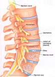
The spinal cord extends from the brain, and travels through the middle part of each vertebra, which is hollow to accommodate the spinal cord and the nerves. Nerves branch out from the spinal cord through openings (foraminae) in the vertebrae, and carry signals between the brain and muscles.
The spinal cord ends between the first and second lumbar vertebrae in the lower back and continues on as nerve roots. This bundle of nerve roots is called the “cauda equina”, as they resemble the tail of a horse. They leave the spinal canal through openings in the vertebrae (foraminae).
Muscles and Ligaments
The muscles and ligaments provide support and stability for your spine and upper body. Strong ligaments, especially those of the lower back, connect your vertebrae and help keep the spinal column in the correct position.
Intervertebral Discs
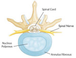
Intervertebral discs (Figure 4) are sandwiched between the vertebrae. They are flat and round, and about a half inch thick. Intervertebral discs are made up of two components. The inner nucleus pulposus is jelly-like and makes up the center of the disc. The jelly is partly made of water and gives the disc flexibility and strength. The outer annulus fibrosus is the flexible outer ring of the disc. It is made up of several layers, similar to elastic bands.
When you are standing or moving, weight is put onto the nucleus. In response, the nucleus expands. The annulus holds the nucleus in place. This allows movement to take place, yet maintains the strength of the spine. In effect, the discs act as shock absorbers for the spine. The most common cause of wear and tear of the spine is the gradual loss of the shock absorbing ability of the disc. The intervertebral disc is a very important structure. Many nerves supply the annulus and, as a result, an injured annulus can cause back pain.
Spinal or Facet Joints
Between the back of the vertebrae are small joints that also help your spine move. At every level of your spine, we have two facet joints (Figure 3) and one intervertebral disc, the so-called three-joint complex. Although the disc does not look like a typical joint, it actually functions like one. The facet joints have a cartilage surface, very much like a hip or a knee joint does. The facet joints are important for allowing rotation of the spine but may develop arthritis and become a source for low back or neck pain.
Looking For A Reliable Neck Orthopaedic Specialist?
Fast Medical Attention, Transparent Fees
Make an appointment for comprehensive care for your neck problems!