Flat Feet in the Paediatric Patient
Flat Foot also known as Pes Planus, describes the altered relationships between the bones of the foot and ankle, and is not a single deformity at a single joint.
These conspire to give the loss of a longitudinal arch, with a heel in exaggerated valgus (swung outwards).

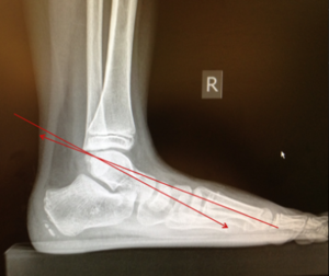
Flat foot in the paediatric patient is not uncommon. This is because children begin life with ligamentous laxity which largely resolves as they reach maturity.
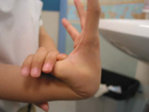
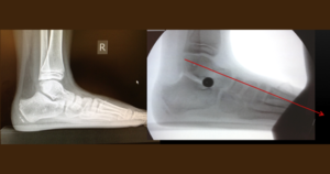
Footprint and Radiological Studies performed by Staheli et al proved that most babies are flat-footed, with the height of the longitudinal arch increasing spontaneously within the 1st decade of life.

Therefore, one must decipher first if it is flexible or rigid, and then whether is it symptomatic or asymptomatic.
What are the Causes and Symptoms of Flat Foot?
A flexible flat foot is one in which the arch is recreated, and the heel swings inwards, upon tip-toeing.
In most cases (approx 66%), the child’s foot is asymptomatic and flexible and the child is able to participate in all activities without complaints.
In approximately a quarter of the cases, the child presents with a flexible and painful flat foot. This could be due to a tight heel cord complex which then cascades into varying subsequent symptoms. 9% of the time, the patient has a rigid flat foot.
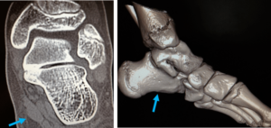
This could be due to tarsal coalition, in which bones of the foot have coalesced to each other to varying extents. As a result of which, the patient can be symptomatic.

How is Flat Foot Treated?
Treatment depends on the type.
The painless asymptomatic flexible flat foot does not require intervention.
The painful flexible flatfoot may benefit from treatment. Treatment starts with the use of insoles and physiotherapy for the stretching of the heel cord.
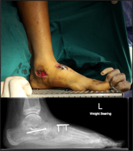
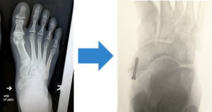
In refractory cases, surgery may be required to address the symptoms. This ranges from the release of the tight heel cord to insertion of an Arthroereisis screw to Bone Cuts and tendon transfers, in order to create a longitudinal arch.
The Arthroereisis screw insertion is based on the premise that a screw is inserted between the bones (sinus tarsi), outside of the joint, in order to allow the developing child’s bones to remodel around it.
This procedure is performed in cases in which the flat foot is mild to moderate in severity, and in which the child is still in the throes of growth.



The rigid flatfoot, when symptomatic, would benefit from the removal of the bridge between the involved bones, so that the joints of the foot can articulate normally without hinder.
These “bridges” can be fibrous, fibre-cartilaginous or bony. It is important to ascertain the type, location and symptoms before proceeding with surgery.
An Os Navicularis syndrome may also be responsible/associated with a painful flat foot. There is an accessory (extra and small) bone next to the navicular bone and within the tibialis posterior tendon complex, which can cause symptoms related to overuse and due to the abnormal excursion of the tendon and pressure on said bone.
While it is largely asymptomatic, cases which are symptomatic may benefit from surgery if conservative methods fail. This involves either fusing the two bones together or excising the bone completely.
Looking For A Reliable Paediatric Orthopaedic Orthopaedic Specialist?
Fast Medical Attention, Transparent Fees
Make an appointment for comprehensive care for your paediatric orthopaedic problems!
